Connective tissues are fundamental components of the body, characterized by a matrix composed of living cells and a non-living substance known as the ground substance. This ground substance itself is comprised of an organic component, typically a protein, and an inorganic component, usually a mineral or water. The principal cell type within connective tissues is the fibroblast, which is responsible for producing the fibers found in nearly all connective tissues. Fibroblasts are motile, capable of mitosis, and can synthesize the specific connective tissue required for a particular function. In addition to fibroblasts, some connective tissues may also contain macrophages, lymphocytes, and, occasionally, leukocytes. Certain tissues possess specialized cells unique to their structure and function.
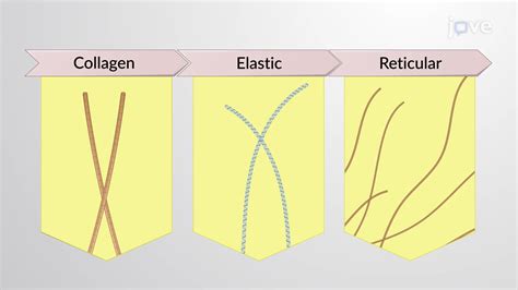
The density of a connective tissue is determined by its matrix. The organic portion of this matrix consists of protein fibers, which can be either collagen, elastic, or reticular fibers. Collagen fibers provide significant strength to tissues, preventing them from tearing or separating from surrounding structures. Elastic fibers, composed of the protein elastin, possess the remarkable ability to stretch up to one and a half times their original length and then return to their original size and shape, imparting flexibility to tissues. Reticular fibers, the third type of protein fiber, are thin strands of collagen that form a supportive network, anchoring the tissue and associated organs.
Types of Connective Tissues and Their Fibers
Loose Connective Tissue
Loose connective tissue, also referred to as areolar connective tissue, is characterized by loosely woven collagen and elastic fibers. It contains a representative sample of all the general components of connective tissue, including fibroblasts and macrophages. In histological preparations, collagen fibers appear relatively wide and stain a light pink, while elastic fibers are thin and stain dark blue to black. The spaces between these formed elements are filled with the matrix, giving the tissue a loose consistency, akin to a pulled-apart cotton ball. Loose connective tissue is found surrounding every blood vessel, helping to maintain its position, and is also present around and between most body organs.
Fibrous Connective Tissues
Fibrous connective tissues are distinguished by a large quantity of collagen fibers and a reduced amount of cells and matrix material. The collagen fibers can be arranged either irregularly, providing resistance to stress from all directions (as seen in the dermis of the skin), or regularly, with strands aligned in parallel for greater tensile strength.
Cartilage
Cartilage is a specialized connective tissue with a substantial amount of matrix and varying amounts of fibers. The cells within cartilage are known as chondrocytes, which are responsible for producing the matrix and fibers. Cartilage is an avascular and aneural tissue that provides structural support and shape to various body parts. Its primary role is to reinforce strategic weight-bearing areas, reducing erosion that would occur from direct articulation of bony surfaces. Cartilage also serves as a major support system for soft tissue structures in the auditory and upper digestive tracts. Due to its avascular nature, cartilage obtains nutrients from the adjacent, well-vascularized perichondrium.
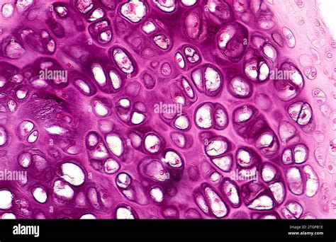
Hyaline Cartilage
Hyaline cartilage is characterized by a matrix containing chondrocytes embedded within it, with relatively few collagen and elastic fibers. This type of cartilage has a glassy appearance when fresh, hence its name (from the Greek word "hyalos" meaning glassy). In H&E stained sections, it appears slightly basophilic overall. Hyaline cartilage contains widely dispersed fine collagen fibers (primarily type II), which contribute to its strength, though these fibers are difficult to visualize in standard sections. It is the most common type of cartilage and is found in the ribs, nose, larynx, and trachea. During embryonic development, hyaline cartilage forms the template for ossification, and remnants persist in the outer nose and movable joints like the knee and shoulder, though it can become damaged with age or trauma.
Elastic Cartilage
Elastic cartilage possesses a large proportion of elastic fibers, which confer significant flexibility. This type of cartilage is found in the ears of most vertebrate animals and in portions of the larynx. While it appears similar to hyaline cartilage in H&E stained sections, special staining is required to visualize the abundant elastic fibers. The chondrocytes in elastic cartilage are situated within a threadlike network of elastic fibers in the matrix. Elastic cartilage provides both strength and elasticity, helping to maintain the shape of certain structures.
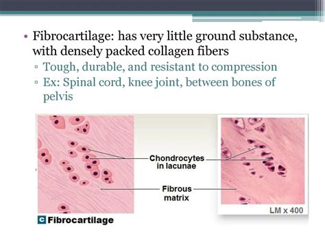
Fibrocartilage
Fibrocartilage is a type of connective tissue notable for its large amount of collagen fibers, which provide tremendous strength. It is a white, densely arranged, opaque tissue with a mixture of both chondrocytes and fibroblasts, often referred to as fibrocartilage cells or fibrochondrocytes. Its composition varies depending on its anatomical location and intended function. Fibrocartilage is considered a transition tissue, blending characteristics of hyaline cartilage and dense fibrous connective tissue. It is the strongest type of cartilage, featuring alternating layers of hyaline cartilage matrix and thick layers of dense collagen fibers oriented along functional stress lines. The collagen fibers are primarily composed of type I collagen, with some type II collagen present. Fibrocartilage is devoid of blood vessels and nerve fibers, relying on the perichondrium for nutrients.
Fibrocartilage is predominantly found in areas subjected to significant mechanical stress, such as the intervertebral discs in vertebrate animals, the menisci of the knee, and the glenoid labrum of the shoulder. In these locations, it acts as a shock absorber and provides structural stability.
The cells within fibrocartilage, particularly in enthesis units, are similar to chondrocytes in their round to oval shape and isolation within lacunae. However, they exhibit limited or no communication among themselves. These cells contain organelles similar to chondrocytes, including lipid droplets, glycogen granules, and intermediate filaments, which likely reinforce the biomechanical properties of the surrounding tissue.
The extracellular matrix of fibrocartilage is a complex mixture contributing to its biomechanical properties. It includes a hydrated, carbohydrate-based ground substance containing proteoglycans with keratin sulphate and chondroitin sulphate side chains, which attract water and contribute to shock absorption. Collagen is a major component, with type I collagen being abundant, a histological feature distinguishing it from other cartilage types. Non-fibrillar collagens, such as types IX and XII, have also been identified in some fibrocartilages.
Fibrocartilage can be classified into several types based on its location and function:
- Intra-articular fibrocartilage: Found in joints allowing flexion and extension, it acts as a buffer, spacer, and thrust pad, enhancing joint stability.
- Connecting fibrocartilage: Located in joints with limited motion, it cushions by spreading compressive forces.
- Stratiform fibrocartilage: Found on joint surfaces where tendons glide over bone, it reduces friction during movement.
- Circumferential fibrocartilage: Ring-shaped, it protects soft tissues in joint margins and improves bone fit within joints.
Bone (Osseous Tissue)
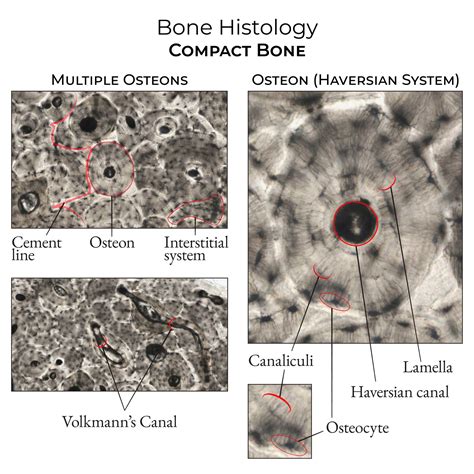
Bone, or osseous tissue, is a specialized connective tissue featuring a substantial amount of two distinct matrix materials. The organic matrix is similar to that found in other connective tissues, containing collagen and elastic fibers that provide strength and flexibility. The inorganic matrix consists of mineral salts, primarily calcium salts, which impart hardness to the tissue. Bone contains three types of cells: osteoblasts (bone-forming cells), osteocytes (mature bone cells found in lacunae), and osteoclasts (bone-resorbing cells involved in remodeling). Bone is categorized into compact bone, characterized by organized units called osteons, and spongy bone, composed of trabeculae.
Adipose Tissue
Adipose tissue, or fat tissue, is considered a connective tissue despite having few fibers and lacking fibroblasts and a substantial matrix. It is composed of adipocytes, cells that store fat in the form of triglycerides for energy metabolism. Adipose tissue also provides insulation to maintain body temperature and cushions organs against damage. Under a microscope, adipocytes often appear empty due to fat extraction during specimen preparation.
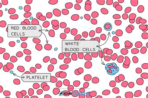
Blood
Blood is classified as a connective tissue because it possesses a matrix, known as plasma, and lacks fibers. The primary living cell types are erythrocytes (red blood cells), responsible for oxygen and carbon dioxide transport, and leukocytes (white blood cells), involved in the immune response. Platelets (thrombocytes) are cell fragments that play a crucial role in blood coagulation to stop bleeding.
Development of Cartilage
Cartilage development, or chondrogenesis, originates from mesenchymal tissue during the fifth gestational week. Signaling molecules such as bone morphogenetic proteins (BMPs) and hedgehog proteins play critical roles in the differentiation of mesenchymal cells into prechondrocytes. These precondrocytes then undergo rapid replication, becoming chondroblasts, and produce the extracellular matrix, leading to their separation into individual lacunae. The process of cartilage differentiation typically occurs centrifugally, with cells at the center maturing before those at the periphery.
The development of fibrocartilage specifically occurs later than other cartilage subtypes, influenced by factors including parathyroid hormone-related peptide and mechanical forces, in addition to genetic involvement. Fibrocartilage is often found in areas like enthesis organs (where tendons attach to bone) and wrap-around regions where tendons change direction around pulley systems.
Disorders Associated with Fibrocartilage
Most disorders involving fibrocartilage are related to traumatic injury. Common examples include:
- Bankart and Hill-Sachs lesions: Associated with shoulder dislocations, these involve damage to the glenoid labrum and humeral head, respectively.
- Acetabular labral tears: Can result from joint laxity, hypermobility, traumatic damage, or femoroacetabular impingement, and can also be influenced by aging and dysplastic changes.
- Intervertebral disc degeneration and herniation: Characterized by the loss of proteoglycans and hydration, leading to increased fibrousness and an inability to distribute stresses, causing pain.
- Triangular Fibrocartilage Complex (TFCC) injuries: Occur at the distal radioulnocarpal joint due to falls on an outstretched hand, torsional forces, or distal radial fractures. These injuries can be classified as traumatic or degenerative.
MRI Anatomy of TFCC
tags: #collagen #fibers #in #fibrocartilage