Patients often express concern when they observe dark spots on spine X-rays, especially when accompanied by severe back pain. It is crucial to understand that these findings require professional interpretation by a healthcare provider in the context of your specific symptoms and medical history.
What Can Cause Dark Spots on Spine X-rays?
The appearance of dark spots on a spine X-ray can be attributed to several factors. It is important to differentiate between normal findings, degenerative changes, and potential pathological conditions.
Normal Findings: Bowel Gas
In many instances, the dark spots you see overlying the spine on an X-ray are simply bowel gas. This is a normal occurrence and does not indicate any underlying spinal pathology.
Degenerative Changes
Radiological reports may describe degenerative changes in the spine, which are common with age. These can manifest as:
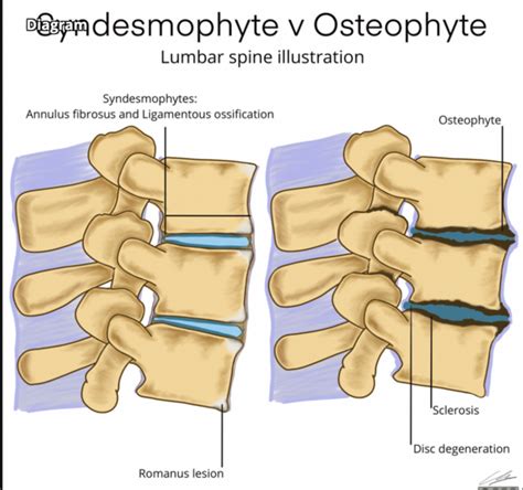
- Osteophyte Formation: This refers to the development of small, excess bone formations. Osteophytes, often called bone spurs, occur naturally over time as bones remodel. They are generally considered a normal part of the aging process.
- End-plate Sclerosis: Sclerosis indicates an increase in bone density, appearing whiter on an X-ray. In the context of spinal end-plates (the top and bottom surfaces of the vertebral bodies), sclerosis can be due to increased weight-bearing or thinning of the cartilage between the bones, potentially contributing to arthritis.
Other Potential Findings
While less common in the context of the spine itself, radiologists may note incidental findings in adjacent structures. For example, a calcific density that appears as a dark spot could potentially represent a kidney stone in a kidney or a gallstone in the gallbladder. These are typically insignificant unless the physician decides further investigation is warranted.
Spondylolisthesis is another condition that may be identified, where one vertebra slips forward over the vertebra below it, most commonly in the lower back.
Understanding Severe Back Pain in Conjunction with X-ray Findings
Severe back pain is a significant symptom that warrants thorough medical evaluation. When combined with X-ray findings, it helps healthcare providers determine the cause and appropriate treatment plan.
Degenerative Disc Disease and Arthritis
Degenerative changes, such as osteophyte formation and end-plate sclerosis, can be associated with conditions like osteoarthritis of the spine. This degenerative joint disease can cause pain, stiffness, and reduced mobility due to the wear and tear on the spinal joints over time.
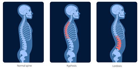
Loss of Normal Curvature
X-rays can also reveal changes in the normal curvature of the spine. For instance, a loss of the normal lumbar lordosis (the inward curve of the lower back) can occur and may be associated with muscle spasms or pain.
Limitations of Spine X-rays
It is important to understand what spine X-rays can and cannot show:
- Bony Structures: X-rays excel at providing detailed images of the bony structures of the spine.
- Soft Tissues: However, X-rays do not show soft tissues well. This includes structures like the cartilage, intervertebral discs, spinal cord, and nerves.
Therefore, X-rays alone are typically insufficient to diagnose conditions affecting soft tissues, such as spinal cord compression or nerve impingement. They also cannot show signs of conditions like Multiple Sclerosis (MS).
When is Further Imaging Recommended?
Given the limitations of X-rays in visualizing soft tissues, further imaging is often recommended to gain a more comprehensive understanding of spinal issues, especially when symptoms are severe or persistent.
Magnetic Resonance Imaging (MRI)
An MRI is generally considered the gold standard for evaluating the soft tissues of the spine. It provides detailed images of the spinal cord, nerves, discs, and ligaments. If X-rays reveal concerning findings or if symptoms suggest a soft tissue issue, your doctor may recommend an MRI.
For example, if you are experiencing neuropathy in your lower extremities and balance issues, and the X-ray shows degenerative changes, an MRI would be beneficial to assess for potential nerve compression or other neurological involvement.
How does an MRI work? | MRI basics explained | Animation
Leukemia and Spine X-rays
While dark spots on a spine X-ray are usually due to bowel gas or degenerative changes, it is important to address the concern about other potential causes, such as leukemia. Leukemia is a cancer of the blood-forming tissues, including bone marrow.
Leukemia and Bone Involvement
In some advanced cases of leukemia, leukemia cells can invade the bone marrow and surrounding tissues of the spine, potentially leading to lesions that might appear as dark spots on an X-ray. This is referred to as extramedullary involvement or spinal lesions.
Leukemia and Skin Symptoms
Leukemia can also manifest with various skin symptoms. These can include:
- Petechiae: Small, pinpoint red or purple spots caused by tiny blood vessel breaks.
- Bruising and Ecchymosis: Discoloration of the skin due to blood leaking into the tissues.
- Leukemia Cutis: Lesions or nodules on the skin caused by the infiltration of leukemia cells.
These skin symptoms can appear on various parts of the body, including the legs, trunk, and back. While these are signs of leukemia, they are distinct from the findings typically seen on spine X-rays, such as bowel gas or degenerative changes.
Diagnosis of Leukemia
Diagnosing leukemia involves a comprehensive evaluation, including:
- Medical History and Physical Examination: Assessing symptoms like fatigue, unexplained weight loss, frequent infections, and bone pain.
- Blood Tests: A complete blood count (CBC) can reveal abnormalities in blood cell levels. An elevated white blood cell count does not always indicate cancer but can be a sign.
- Bone Marrow Biopsy: This is the definitive test for diagnosing leukemia, where a sample of bone marrow is examined for leukemia cells.
- Skin Biopsy: If leukemia cutis is suspected, a biopsy of skin lesions may be performed.
It is crucial to remember that the interpretation of spine X-rays should be done by a qualified radiologist and discussed with your physician. If there is a suspicion of leukemia or other serious conditions, your doctor will guide you through the necessary diagnostic steps.
Understanding X-ray Technology
X-rays utilize invisible electromagnetic energy beams to create images of internal tissues, bones, and organs. Here's a basic explanation of how they work:
- Radiation Penetration: When X-rays pass through the body, different tissues absorb varying amounts of radiation.
- Bone Density: Dense structures like bones absorb more X-rays and appear white on the image because fewer beams pass through them.
- Soft Tissues: Softer tissues, such as muscles, fat, and organs, allow more X-rays to pass through, resulting in darker gray or black areas on the image.
- Bowel Gas: Air within the digestive tract (bowel gas) allows almost all X-rays to pass through, appearing very dark on the film.
- Fractures: A break or fracture in a bone is less dense than the surrounding bone, allowing more X-rays to pass through, which appears as a dark line within the white bone.

Preparation and Procedure for Spine X-rays
Preparing for and undergoing a spine X-ray is generally straightforward:
- Preparation: Typically, no special preparation is needed. You can eat and drink normally. You may be asked to wear comfortable clothing and remove jewelry or metal objects that could interfere with the images.
- Procedure: You will be positioned on an X-ray table, either lying down or standing, depending on the specific views required. A lead apron may be placed over areas not being examined to minimize radiation exposure. The technologist will step behind a protective barrier while taking the images. You may be asked to hold your breath or remain still for a few moments during the exposure.
- Duration: The procedure usually takes about 15 minutes, though it may vary depending on the number of images needed.
- Pain: X-rays themselves are painless. However, if you have a painful injury or condition, the positioning for the X-ray might cause temporary discomfort.
- Risks: X-rays use a small amount of radiation. While occasional medical X-rays are generally safe, it's important to inform your healthcare provider if you are pregnant or have had many X-rays in the past.