Immunoglobulins, also known as antibodies, are crucial proteins produced by plasma cells as part of the humoral immune response. This response is triggered when the immune system encounters antigens. Following the initial contact with an antigen, the immune system first produces antibodies of the IgM class. Subsequently, antibodies of the IgG and IgA classes are formed.
Quantitative determination of immunoglobulins provides valuable insights into the humoral immune status. In cases of monoclonal immunoglobulin proliferations, conditions such as plasmacytomas, Waldenstrom disease, and heavy-chain disease may be indicated. Monoclonal immunoglobulinemia necessitates thorough differential diagnostic investigations beyond simple quantitative measurement.
Local immune reactions can lead to elevated immunoglobulin levels, particularly IgG, within the cerebrospinal fluid (CSF). Conversely, IgA levels may increase during asparaginase treatment, pregnancy, exercise, and in individuals with alcohol use disorder. IgA levels can decrease with prolonged exposure to benzene and after a year of abstinence from alcohol. Certain medications and substances, including diphenylhydantoin, dextran, methyl prednisolone, toluene, xylol, and oral contraceptives, may also lower IgA levels.
The gamma globulin band observed in conventional serum protein electrophoresis comprises all five major classes of immunoglobulins. Elevations in IgG can result from polyclonal immunoglobulin production, indicating a widespread immune response.

Understanding IgG Antibodies
IgG is the most abundant circulating immunoglobulin, found in both plasma and interstitial fluid. It possesses a monomeric structure and is classified into four subclasses: IgG1, IgG2, IgG3, and IgG4. A deficiency in any of these IgG subclasses can occur but may be overlooked if the dominant IgG1 levels remain within the normal range.
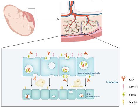
Understanding the age-specific appearance of IgG subclasses is essential for comprehending the immune responses in very young individuals. It is noteworthy that IgA deficiency can be associated with deficiencies in IgG2 and IgG4. IgG is unique among immunoglobulins for its ability to cross the placenta in significant amounts. Consequently, any IgG-mediated disease can be temporarily transmitted to the fetus, with Graves' disease serving as a classic example.
Functions of IgG
IgG antibodies play a vital role in responding to pathogens through several mechanisms:
- Alerting other immune cells: IgG complexes can bind to pathogens, signaling other immune cells to destroy them. This is achieved through opsonization (coating the pathogen to attract immune cells) or by activating the complement system, a cascade of proteins that damage or destroy the pathogen.
- Neutralizing pathogens: When an IgG antibody binds to a pathogen, it can prevent the pathogen from attaching to and invading the body's cells.
Production of IgG
The production of IgG is part of the immune system's response to encountering a potentially harmful antigen. Depending on whether the immune system has previously encountered the pathogen, this leads to either a primary or secondary immune response. During the primary response, the body initially produces IgM antibodies, which help combat the intruder. Subsequently, the immune system shifts to producing IgG antibodies, which are more widely distributed and possess enhanced capabilities for fighting infections.
Following the initial encounter, the immune system generates memory cells. Upon re-exposure to the same pathogen, the body can more rapidly switch to producing IgG antibodies, which are specifically tailored to recognize and neutralize the invader. Vaccines are designed to stimulate this memory response, enabling the body to produce IgG antibodies against specific pathogens without causing illness.
Distribution of IgG in the Body
IgG is the most prevalent antibody in blood and the fluid surrounding cells. Its small size facilitates easy movement into tissues throughout the body. Notably, IgG is the only antibody capable of crossing the placenta, conferring temporary immune protection to newborns for approximately six months after birth.
Elevated IgG Levels: Causes and Associated Conditions
Elevated IgG levels (typically above 16 g/L) can be indicative of several conditions, including chronic active infections, inflammation, or plasma cell disorders. The specific causes of a raised IgG level can include:
- Chronic active infection
- Chronic liver disease (e.g., cirrhosis)
- Sarcoidosis
- Autoimmune diseases
- Parasitic diseases
- Multiple myeloma (a type of cancer affecting plasma cells)
In contrast, low IgG levels can be associated with acquired or hereditary immunodeficiency, pregnancy, protein-losing syndromes, Waldenstrom's macroglobulinemia, and non-IgG myeloma.
Cerebrospinal Fluid (CSF) IgG Index
CSF, a clear, colorless fluid that circulates within and around the brain and spinal cord, plays a crucial role in cushioning the central nervous system. Normally, only a small amount of IgG is present in the CSF. Elevated levels of IgG in the CSF may suggest an infection, inflammation, or autoimmune disease affecting the central nervous system.
The CSF IgG index is a diagnostic tool used to assess diseases of the central nervous system, frequently employed in the diagnosis of multiple sclerosis (MS). A majority of individuals with MS exhibit higher-than-normal IgG levels in their CSF. A CSF IgG index may be recommended for individuals experiencing symptoms of a central nervous system disorder, particularly MS.
Lumbar Puncture: Everything You Need to Know
Symptoms of Central Nervous System Disorders
The symptoms associated with central nervous system disorders, such as multiple sclerosis, can be highly variable. They may manifest as intermittent episodes or a steady progression of worsening symptoms.
Lumbar Puncture for CSF Analysis
To obtain a sample of cerebrospinal fluid, a medical provider performs a procedure called a spinal tap, also known as a lumbar puncture. This procedure is typically conducted in a hospital setting. The provider will first clean the patient's back and administer a local anesthetic to numb the area, minimizing discomfort during the procedure.
Once the area is completely numb, a thin, hollow needle is inserted between two vertebrae in the lower spine. A small amount of CSF is then withdrawn for laboratory testing. After the procedure, the patient may be asked to lie flat for an hour or two. The risks associated with a spinal tap are minimal, usually involving a brief sensation of a pinch or pressure during needle insertion, potential minor bleeding at the puncture site, or a headache that can last from several hours to over a week.
Interpreting CSF IgG Index Results
A CSF IgG index that is lower than normal might indicate an impaired ability of the body to produce sufficient IgG antibodies, potentially increasing susceptibility to infections. The exact causes of this deficiency are not yet fully understood by experts.