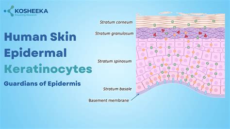
The outermost layer of human skin, the epidermis, is primarily composed of specialized cells known as keratinocytes. These cells constitute the largest percentage of the epidermis, making up approximately 90% of all epidermal cells. Each keratinocyte originates from the deepest part of the epidermis and gradually migrates upwards through its various layers. During this journey, the cell undergoes a transformation, changing its shape and becoming progressively tougher as it fills with a robust protein called keratin. This protein is instrumental in providing strength and elasticity to the skin, as well as aiding in water retention.
The Multifunctional Role of Keratinocytes
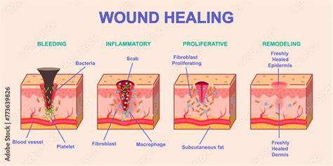
Beyond their primary function of forming a protective barrier, keratinocytes are actively involved in several crucial processes. They play a significant role in wound healing; upon experiencing a minor cut or injury, keratinocytes rapidly move to cover the damaged area, facilitating the restoration of the skin to its original state. Furthermore, keratinocytes act as sentinels for the immune system. When they detect the presence of harmful substances like bacteria or chemicals, they emit signals that alert other immune cells, prompting a swift response. A healthy population of keratinocytes is essential for maintaining skin that is strong, smooth, and radiant, with new active layers constantly being introduced from below as old cells are replaced.
Understanding Keratinocytes: Structure and Function
To fully appreciate how keratinocytes function, it is helpful to define them as specialized skin cells responsible for producing keratin, a strong, flexible protein that provides structural integrity to the skin's outer layer, as well as to hair and nails. At any given moment, each keratinocyte is engaged in maintaining healthy and stable skin. These cells are interconnected by robust links, described as cellular 'glue,' which hold the skin intact while allowing for significant stretching and movement without damage. Internally, each keratinocyte operates like a small factory, manufacturing keratin fibers, enzymes, and lipids vital for skin strength and moisture content. The lipids secreted by keratinocytes form natural oils that help retain moisture, preventing dryness and flaking. Keratinocytes also engage in communication with other skin cells to maintain balance, sending out instructions that regulate skin growth, healing, and responses to injury.
A remarkable characteristic of keratinocytes is their ability to self-renew, a process that dictates the life cycle of skin cells. New keratinocytes are continuously formed deep within the basal layer. As they ascend through the skin's layers, they alter their shape and structure. Eventually, cells on the surface die and naturally shed through exfoliation, making way for new cells. This continuous renewal process keeps the skin fresh, taut, and smooth, representing a simple yet extraordinary biological mechanism. In fact, the entire top layer of skin is replaced by new keratinocytes every few weeks.
Layers of the Epidermis and Keratinocyte Differentiation
Keratinocytes are not uniform; they exhibit variations depending on their location within the epidermis. The basal layer is where keratinocytes originate and form the deepest layer of the epidermis. Moving upwards, the next type of keratinocyte is found in the spinous layer, also known as the stratum spinosum or prickle cell layer. The uppermost layer, the stratum corneum, contains keratinocytes that are no longer alive. In this layer, they become flattened, hardened plates densely packed with keratin, serving as a protective shield for the body.
The life of a skin cell begins at the bottom of the epidermis, specifically in the basal layer, which is dedicated to cell production. Here, young, active keratinocytes are created and undergo division to produce more cells. As these keratinocytes ascend, their shape changes from round and soft to flattened, and they begin to accumulate keratin, a tough protein that imparts hardness and resistance to damage. The entire journey from the basal layer to the skin's surface typically takes about one month. During this process, the keratinocyte becomes increasingly specialized, packing itself with a dense structure that locks in moisture and forms a barrier against toxic substances. By the time a keratinocyte reaches the stratum corneum, it is dead. These flattened, dead keratinocytes are tightly packed, forming a robust protective layer. Even in death, these cells continue to serve an important protective function before being naturally shed through a process called desquamation. This gentle, continuous shedding allows new cells to emerge from beneath.
Key Functions: Protection and Repair
The primary role of keratinocytes is protection. They act as a defensive barrier against external threats entering the human body. The keratin they produce forms a protective shield, maintaining the skin's thickness to withstand daily wear and tear. This barrier also helps prevent excessive water loss, thereby averting dehydration. Keratinocytes are responsive to environmental stimuli, such as sunlight. Upon exposure to UV radiation, they signal for melanin production. In cases of skin injury, keratinocytes migrate to the affected site to collaborate with other cell types in forming new tissue and filling the wound, thus providing a barrier against infection and aiding in healing. Keratinocytes respond rapidly to repair skin damage from cuts, burns, and everyday stressors.
Maintaining Keratinocyte Health
Healthy skin is intrinsically linked to the health of its keratinocytes. Factors such as excessive sun exposure, pollution, and harsh chemicals can damage these cells, leading to dryness, flaking, and sensitivity. Maintaining strong keratinocytes relies on good skincare practices, including keeping the skin clean and well-moisturized to support its barrier function. Dietary intake rich in vitamins and adequate water consumption also contribute to their health. Scientists actively study dead keratinocytes to explore potential therapeutic improvements for skin conditions.
Keratinocytes in Scientific Research and Biotechnology
Keratinocytes are considered one of the most valuable cell lines in scientific research, significantly contributing to our understanding of skin biology. Researchers utilize keratinocytes to investigate the effects of new compounds and treatments on the skin. Comprehending keratinocyte function aids in developing strategies to enhance skin injury healing and improve protection against various forms of damage. Keratinocytes play a critical role in studies examining the immune response to skin infections or irritation. Furthermore, biotechnologists are developing keratinocyte cultures for creating safe and effective products for research and healthcare applications.
The dynamic life of keratinocytes underscores the skin's remarkable ability to remain alive, strong, and protective. These cells tirelessly work to shield the body from harm, repair damage, and maintain hydration. Understanding the nature, function, and life cycle of keratinocytes reveals the intricate complexity of the skin as an organ. The foundation of skin health begins with these fundamental cellular units.
AP1: SKIN: KERATINIZATION IN EPIDERMIS
Keratin: The Structural Protein
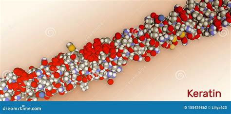
Keratin is a family of structural fibrous proteins, also known as scleroproteins. It serves as the key structural material for scales, hair, nails, feathers, horns, claws, hooves, and the outer layer of skin in vertebrates. Keratin also protects epithelial cells from damage and stress. It is highly insoluble in water and organic solvents.
Types of Keratin
Alpha-keratins (α-keratins) are found in all vertebrates and form structures like hair, the outer layer of skin, horns, nails, claws, and hooves in mammals, as well as the slime threads of hagfish. The baleen plates of filter-feeding whales are also made of keratin. Keratin filaments are abundant in keratinocytes of the hornified epidermis, representing proteins that have undergone keratinization. They are also present in epithelial cells generally.
Beta-keratins (β-keratins) are found exclusively in sauropsids (reptiles and birds). They constitute nails, scales, and claws of reptiles, some reptile shells, and the feathers, beaks, and claws of birds. These keratins are primarily formed in beta sheets and are structurally and genetically distinct from α-keratins.
Keratin Structure and Composition
Keratins, also referred to as cytokeratins, are polymers of type I and type II intermediate filaments found in chordates. The human genome encodes 54 functional keratin genes. A distinguishing feature of keratins is the presence of significant amounts of the sulfur-containing amino acid cysteine. Cysteine residues form disulfide bridges, which provide additional strength and rigidity through permanent, thermally stable crosslinking, similar to how sulfur bridges stabilize vulcanized rubber. Human hair, for example, is approximately 14% cysteine. The pungent smell of burning hair and skin is attributed to volatile sulfur compounds formed during this process.
More flexible and elastic keratins, such as those in hair, have fewer interchain disulfide bridges compared to the keratins in harder structures like fingernails, hooves, and claws. These harder keratins exhibit greater similarity to their analogs in other vertebrate classes. Hair and other α-keratins are composed of α-helically coiled single protein strands, which further twist into superhelical ropes that can be coiled. Keratins are categorized into 'hard' and 'soft' forms, or 'cytokeratins' and 'other keratins'.
Keratin and the Cytoskeleton
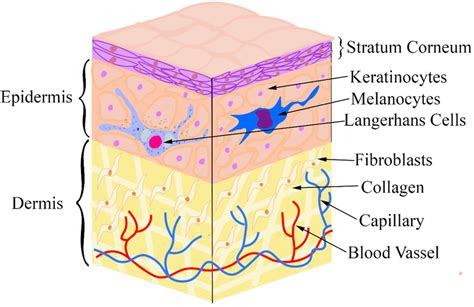
In many cell types, including those in the dermis, keratin filaments and other intermediate filaments function as part of the cytoskeleton, providing mechanical stability to cells against physical stress. Cells in the epidermis contain a structural matrix of keratin, making the outermost skin layer nearly waterproof. Along with collagen and elastin, keratin contributes to the skin's strength. Physical stimuli like rubbing and pressure can lead to thickening of the outer, cornified layer of the epidermis, forming protective calluses, which are beneficial for athletes and musicians.
Keratin in Disease and Research
Keratin expression is a useful marker in determining the epithelial origin of anaplastic cancers. Tumors that express keratin include carcinomas, thymomas, sarcomas, and trophoblastic neoplasms. The specific expression pattern of keratin subtypes can aid in predicting the primary tumor's origin when assessing metastases.