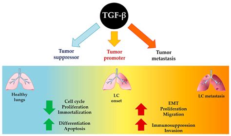
Uveal melanoma (UM) is a severe form of cancer characterized by limited therapeutic options. In advanced stages, UM cells predominantly metastasize to the liver, presenting a significant challenge for effective treatment of these metastatic lesions. Transforming growth factor beta (TGF-β) plays a dual role in cancer progression. Initially, it can inhibit tumor growth, but at later stages, it promotes metastasis, often by inducing an epithelial-to-mesenchymal transition (EMT). This suggests that targeting the TGF-β pathway could be a promising strategy for treating metastatic UM.
Characterization of UM Cell Lines
To investigate the potential of targeting the TGF-β pathway in UM, this study focused on characterizing the Mel270 and 92.1 cell lines. These cell lines were identified as exhibiting a pseudoepithelial/mesenchymal phenotype.
Phenotypic Analysis
The research first aimed to characterize the pseudoepithelial/mesenchymal phenotype of the UM cell lines Mel270 and 92.1. The results demonstrated that these UM cell lines indeed exhibited a mesenchymal phenotype.
Response to Transforming Growth Factor Beta (TGF-β)
Following phenotypic characterization, the study proceeded to evaluate the responsiveness of the Mel270 and 92.1 cell lines to TGF-β treatment. This evaluation was conducted in vitro to understand the functional impact of TGF-β on their cell viability.
Cytostatic Effects of TGF-β
The cell lines were treated with TGF-β to assess their reaction to the cytokine. The findings indicated that TGF-β exerted a cytostatic effect on the UM cell lines. This means that TGF-β treatment led to a halt in cell proliferation rather than cell death.
Summary of Findings
The study concluded with two key findings: Firstly, the uveal melanoma cell lines Mel270 and 92.1 possess a mesenchymal phenotype and respond to TGF-β treatment in vitro. Secondly, TGF-β promotes a cytostatic effect on these UM cell lines.
Related Research and Cell Line Information
Previous research has explored various aspects of uveal melanoma, including the expression of MAGE genes during disease progression, molecular cytogenetic evaluations of UM cell lines, and comprehensive genetic and molecular characterization of UM cell lines. These studies contribute to a broader understanding of the molecular underpinnings of UM and the characteristics of established cell line models.
- Expression of MAGE genes in ocular melanoma during progression from primary to metastatic disease (Chen et al., 1997).
- Molecular cytogenetic evaluation of 10 uveal melanoma cell lines (White et al., 2006).
- Genetic and molecular characterization of uveal melanoma cell lines (Griewank et al., 2012).
- A high-throughput panel for identifying clinically relevant mutation profiles in melanoma (Dutton-Regester et al., 2012).
- Evidence of epidermal growth factor receptor expression in uveal melanoma: inhibition of epidermal growth factor-mediated signalling by gefitinib and cetuximab triggered antibody-dependent cellular cytotoxicity (Amaro et al., 2013).
- Uveal melanoma cell lines: where do they come from? (Jager et al., 2016).
- Protein and mRNA expression in uveal melanoma cell lines are related to GNA and BAP1 mutation status (Gelmi et al., 2024).