Regularly examining your skin is a crucial step in detecting potential skin cancers. It involves looking for any new moles or lesions, as well as changes in existing moles. When examining moles, the ABCDEs of melanoma serve as a helpful guide.
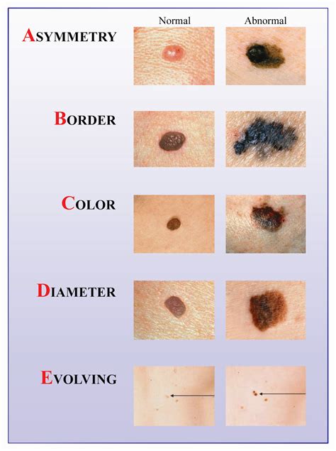
The ABCDEs of Melanoma
Melanomas can exhibit various characteristics, and the ABCDE rule is designed to help identify suspicious lesions:
- A is for Asymmetry: Normally, a spot on our skin is symmetrical. However, with melanoma, one half may look different than the other.
- B is for Border: Changes in the border of a mole or spot can be a sign of melanoma. The edges may be irregular, notched, or blurred.
- C is for Color: Melanomas often contain multiple shades of brown or black but can sometimes be mixed with white, gray, blue, or red. These colors are typically unevenly or irregularly distributed.
- D is for Diameter: Melanoma spots are typically large and greater than 6 mm (1/4 inch) in diameter.
- E is for Evolving: This refers to any changes in the size, color, shape, or structure of a spot over time. A nodular melanoma, for instance, tends to grow rapidly with changes noted over days or weeks.
Additionally, the "Ugly Duckling" sign is important to remember. This refers to a mole or lesion that is unlike the others on your skin. It might be a lighter or darker colored mole or lesion than the others around it, or it might be a larger or smaller mole or lesion than others around it.
How to Perform a Skin Self-Examination
Performing a skin self-examination at home is a simple yet powerful habit. You will need a well-lit room, a full-length mirror, and a hand mirror.
Steps for Skin Self-Examination:
- Front and Back: Use the full-length mirror to examine your entire front and back.
- Sides: Raise your arms and look at the left and right sides of your body.
- Face and Scalp: Check your lips, tongue, inner cheeks, and use both mirrors to inspect the back of your scalp and neck. Part your hair or use a blow dryer for a closer look. You can also ask your hairdresser or barber to point out any unusual spots.
- Arms and Hands: With your elbows bent, check your forearms, upper arms, palms, and between your fingers.
- Torso and Genital Area: Use mirrors to look at your back, buttocks, and genital area.
- Breasts: If you have breasts, lift them to see the skin underneath.
- Legs and Feet: Sit down if needed. Check the backs of your legs, soles of your feet, and between your toes.
During your self-exam, note any moles, blemishes, or birthmarks from head to toe. Use a small ruler to measure their size, and watch for changes in color, shape, or size, or any sore that doesn’t heal. Taking photos can help you track changes over time.
Frequency of Examination
Check your skin about once a month, ideally after a bath or shower. Keep track of any spots or changes and where they appear. Contact your healthcare provider if you notice anything new or changing.
Understanding Skin Cancer Terminology
A comprehensive understanding of medical terms is essential in discussing and treating skin cancer. Here are some key definitions:
Common Skin Conditions and Growths:
- Actinic keratosis (ak-TIH-nik KAYR-uh-TOH-sis): A thick, scaly patch of skin that may become cancer.
- Benign (beh-NINE): Not cancerous. Benign tumors may grow larger but do not spread to other parts of the body.
- Bowen disease (BOH-en dih-ZEEZ): A skin disease marked by scaly or thickened patches on the skin, often caused by prolonged exposure to arsenic. These patches may become malignant.
- Carcinoma in situ (KAR-sih-NOH-muh in SY-too): A group of abnormal cells that remain in the place where they first formed and have not spread. These abnormal cells may become cancer.
- Dysplastic nevus (dis-PLAS-tik NEE-vus): A type of nevus (mole) that looks different from a common mole, often being larger with less defined borders and uneven color.
- Melanoma (MEH-luh-NOH-muh): A form of cancer that begins in melanocytes (cells that make the pigment melanin).
- Melanoma in situ (MEH-luh-NOH-muh in SY-too): Abnormal melanocytes are found in the epidermis (outer layer of the skin) but have not yet spread.
- Merkel cell carcinoma (MER-kel sel KAR-sih-NOH-muh): A rare type of cancer that forms on or just beneath the skin, usually in sun-exposed areas.
- Mole: A benign (not cancer) growth on the skin formed by a cluster of melanocytes.
- Nevoid basal cell carcinoma syndrome (NEE-voyd BAY-sul SEL KAR-sih-NOH-muh SIN-drome): A genetic condition that increases the risk of basal cell carcinoma.
- Squamous cell (SKWAY-mus sel): Flat cells that cover inside and outside surfaces of the body.
- Tumor (TOO-mer): An abnormal mass of tissue resulting from uncontrolled cell division or a failure of cells to die.
Diagnostic and Treatment Procedures:
- Biopsy (BY-op-see): The removal of cells or tissues for examination by a pathologist. Types include incisional biopsy (sample removed), excisional biopsy (entire lump removed), and needle biopsy (sample removed with a needle).
- Cryosurgery (KRY-oh-SER-juh-ree): A procedure where tissue is frozen to destroy abnormal cells.
- CT scan: A series of detailed X-ray pictures taken from different angles by a computer.
- Excisional biopsy (ek-SIH-zhun-al BY-op-see): A surgical procedure to remove an entire lump or suspicious area for diagnosis.
- Excisional skin surgery (ek-SIH-zhun-al SER-juhree): Surgical removal of moles, cysts, skin cancer, and other skin growths.
- Fine-needle aspiration biopsy (AS-pih-RAY-shun BY-op-see): Removal of tissue or fluid with a thin needle for examination.
- Mohs surgery (MOZE SER-juh-ree): A surgical procedure for skin cancer where individual layers of cancer tissue are removed and examined under a microscope one at a time.
- PET scan: A procedure using radioactive glucose to create pictures of areas inside the body where glucose is used, helping to find cancer cells.
- Punch biopsy (BY-op-see): Removal of a small disk-shaped sample of tissue using a sharp, hollow device.
- Radiation therapy (RAY-dee-AY-shun THAYR-uhpee): The use of high-energy radiation to kill cancer cells and shrink tumors.
- Shave biopsy (BY-op-see): Removal of a skin abnormality and a thin layer of surrounding skin with a small blade for examination.
- Sentinel lymph node biopsy (limf node): Removal and examination of the first lymph node(s) to which cancer cells are likely to spread.
Medical and Biological Terms:
- Biological therapy (BY-oh-LAH-jih-kul THAYR-uhpee): Treatment to boost or restore the immune system's ability to fight cancer.
- Blood vessel: A tube through which blood circulates in the body.
- Cell (sel): The individual unit that makes up the tissues of the body.
- Clinical trial (KLIH-nih-kul TRY-ul): A research study testing new medical approaches in people.
- Dermis (DER-mis): The inner layer of the two main layers of the skin.
- Fluorouracil (floor-oh-YOOR-uh-sil): A drug used to treat certain cancers and skin conditions.
- Human papillomavirus (HYOO-mun PA-pih-LOH-muh-VY-rus): A virus that can cause abnormal tissue growth and, in some cases, cervical cancer.
- Imiquimod (ih-MIH-kwee-mod): A drug used to treat early basal cell skin cancer and other skin conditions.
- Incisional biopsy (in-SIH-zhun-al BY-op-see): A surgical procedure where a portion of a lump or suspicious area is removed for diagnosis.
- Infection: Invasion and multiplication of germs in the body.
- Inflammation (IN-fluh-MAY-shun): Redness, swelling, pain, and/or a feeling of heat in an area of the body.
- Interferon (in-ter-FEER-on): A biological response modifier that can slow tumor growth.
- Interleukin-2 (in-ter-LOO-kin): A protein that boosts the immune system in cancer therapy.
- Intravenous (IN-truh-VEE-nus): Into or within a vein.
- Local anesthesia (LOH-kul A-nes-THEE-zhuh): Temporary loss of feeling in a small area of the body.
- Lymph node (limf node): Rounded mass of lymphatic tissue that filters lymph and stores lymphocytes.
- Lymph vessel (limf): A thin tube that carries lymph through the lymphatic system.
- Malignant (muh-LIG-nunt): Cancerous.
- Margin: The edge or border of tissue removed in cancer surgery.
- Medical oncologist (MEH-dih-kul on-KAH-loh-jist): A doctor specializing in diagnosing and treating cancer.
- Metastasis (meh-TAS-tuh-sis): The spread of cancer from one part of the body to another.
- MRI: Magnetic Resonance Imaging, creating detailed pictures of areas inside the body.
- Organ: A part of the body that performs a specific function.
- Photodynamic therapy (FOH-toh-dy-NA-mik THAYR-uh-pee): Treatment with drugs that become active when exposed to light.
- Radiation (RAY-dee-AY-shun): Energy released in the form of particle or electromagnetic waves.
- Registered dietitian (dy-eh-TIH-shun): A health professional specializing in diet and nutrition.
- Risk factor: Something that increases the chance of developing a disease.
- Side effect: A problem that occurs when treatment affects healthy tissues or organs.
- Supportive care: Care given to improve the quality of life for patients with serious diseases.
- Surgery (SER-juh-ree): A procedure to remove or repair a part of the body.
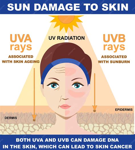
- Ultraviolet radiation (UL-truh-VY-oh-let RAY-deeAY-shun): Invisible rays from the sun, sun lamps, and tanning beds that can damage the skin.
- UVA radiation (RAY-dee-AY-shun): A type of ultraviolet radiation that penetrates deeper into the skin.
- UVB radiation (RAY-dee-AY-shun): A type of ultraviolet radiation that causes sunburn.
- Vaccine (vak-SEEN): A substance meant to cause the immune system to respond to a tumor or microorganisms.
- Vitamin D (VY-tuh-min): A nutrient essential for bone health, which the skin can produce when exposed to sunshine.
- X-ray: A type of high-energy radiation used to diagnose diseases by making pictures of the inside of the body.
- Xeroderma pigmentosum (ZEER-oh-DER-ma pigmen-TOH-sum): A genetic condition with extreme sensitivity to ultraviolet radiation.
Skin Biopsy Explained: A 3D Visualization of Punch, Shave & Excision Techniques
The Importance of Early Detection
Catching skin cancer in its early stages provides more treatment options and a better chance of successful outcomes. While not every skin change is cancer, and not every lesion fitting the ABCDE or ugly duckling criteria is skin cancer, it's crucial to trust your instincts. If something doesn't look or feel right, reach out to your healthcare provider.
Scientists have long thought that UVB radiation can cause melanoma and other types of skin cancer. They now think that UVA radiation also may add to skin damage that can lead to skin cancer and cause premature aging. For this reason, skin specialists recommend that people use sunscreens that reflect, absorb, or scatter both kinds of ultraviolet radiation.
Abbreviations in Dermatology
Abbreviations are commonly used in healthcare practices to save time when documenting patient information. However, the use of non-standard abbreviations can lead to confusion, especially with a growing and diverse healthcare workforce. To mitigate these risks, practices can implement the following strategies:
- A practice-specific glossary of abbreviations: This list should be kept up to date and easily accessible to all staff. A dermatology-specific Electronic Health Record (EHR) can be a valuable tool for maintaining this list in real-time.
- A "never use" list of potentially confusing abbreviations: Practices that adhere to avoiding specific abbreviations significantly reduce confusion.
- Regular staff training: Educate staff on the dangers of using ambiguous and non-standard abbreviations.
- Regular document audits: Conduct spot checks of practice documents to identify ambiguous abbreviations.
A dermatology-specific EHR can further reduce the risks of confusing abbreviations by offering customizable templates pre-populated with common diagnoses, procedures, and treatment plans, thus eliminating the need for extensive abbreviation.